
PUBLICATIONS
Independent work at Univ of Guelph :
Vahidi Lab trainees and staff scientists are underlined.
M. L. Nosella, T. H. Kim, S. K. Huang, R. W. Harkness, M. M. Goncalves, A. Pan, M. Tereshchenko, S. Vahidi, J. L. Rubinstein, H. O. Lee, J. D. Forman-Kay, L. E. Kay, “Poly-ADP-ribosylation enhances nucleosome dynamics and organizes DNA damage repair components within biomolecular condensates”, Molecular Cell, 84 (3), 429-446 (2024).

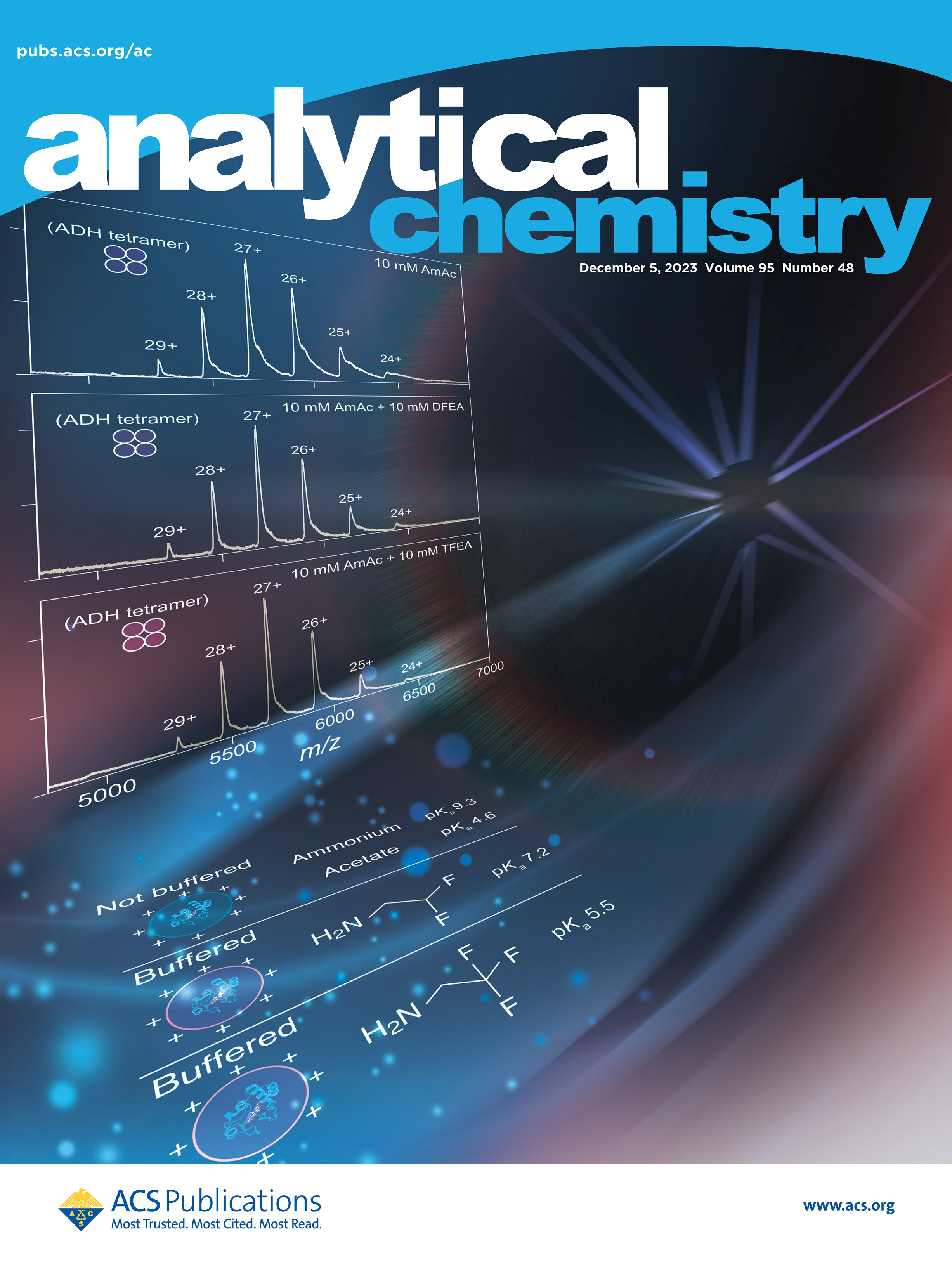
B. T. V. Davis, A. Velyvis*, and S. Vahidi*, “Fluorinated ethylamines as electrospray-compatible neutral pH buffers for native mass spectrometry”, Analytical Chemistry, 95 (48) 17525-17532 (2023). ACS Editors’ Choice and Cover Feature, U of G news release * corresponding author

J. M. Di Trani, A. A. Gheorghita, M. Turner, P. Brzezinski, P. Ädelroth, S. Vahidi, P. L. Howell, and J. L. Rubinstein, “Structure of the bc1-cbb3 respiratory supercomplex from Pseudomonas aeruginosa” Proc. Natl. Acad. Sci. U.S.A. 120 (40) e2307093120 (2023).

Y. Liang, A. Plourde, S. A. Bueler, J. Liu, P. Brzezinski*, S. Vahidi*, and J. L. Rubinstein*, “Structure of mycobacterial respiratory Complex I” Proc. Natl. Acad. Sci. U.S.A. 120 (13) e2214949120 (2023).* corresponding author
See SickKids Hospital and U of Guelph news release.

Graduate and postdoctoral work:
Z. A. Ripstein*, S. Vahidi*, J. L. Rubinstein, and L. E. Kay, “A pH-Dependent Conformational Switch Controls N. meningitidis ClpP Protease Function” J. Am. Chem. Soc. 142, 20519–20523 (2020). * Co-first authorship and corresponding author

S. Vahidi*, Z. A. Ripstein*, J. B. Juravsky, E. Rennella, A. L Goldberg, A. K. Mittermaier, J. L. Rubinstein, and L. E. Kay, “An allosteric switch regulates Mycobacterium tuberculosis ClpP1P2 protease function as established by cryo-EM and methyl-TROSY NMR” Proc. Natl. Acad. Sci. U.S.A. 117, 5895-5906 (2020). * Co-first authorship.

Read commentary by Dr. Andrew Byrd, Structural Biophysics Laboratory, National Cancer Institute, Frederick, MD, USA, in PNAS “Could confounding the allosteric communication of biotic machinery be an alternative path to antibiotics?” DOI: 10.1073/pnas.2002666117
Z. A. Ripstein*, S. Vahidi*, W. A. Houry, J. L. Rubinstein, and L. E. Kay, “A processive rotary mechanism couples substrate unfolding and proteolysis in the ClpXP degradation machinery” eLife 9:e52158, DOI: 10.7554/eLife.52158 (2020). * Co-first authorship and corresponding author

Read Insight Article by Profs. Francis TF Tsai and Christopher Hill, Baylor College of Medicine & University of Utah School of Medicine, US in eLife “Protein Unfolding: Same structure, different mechanisms?” DOI: 10.7554/eLife.56501
M. F. Mabanglo*, E. Leung*, S. Vahidi*, T. V. Seraphim*, B. T. Eger, S. Bryson, V. Bhandari, J. L. Zhou, Y. Mao, K. Rizzolo, M. M Barghash, J. D Goodreid, S. Phanse, M. Babu, L. RS. Barbosa, C. H. Ramos, R. A. Batey, L. E. Kay, E. F. Pai, W. A. Houry, “ClpP protease activation results from the reorganization of the electrostatic interaction networks at the entrance pores” Commun. Biol. 2, 410 (2019). * Co-first authorship.

S. Vahidi*, Z. A. Ripstein*, M. Bonomi, T. Yuwen, M. F. Mabanglo, J. B. Juravsky, K. Rizzolo, A. Velyvis, W. A. Houry, M. Vendruscolo, J. L. Rubinstein, and L. E. Kay, “Reversible inhibition of the ClpP protease via an N-terminal conformational switch” Proc. Natl. Acad. Sci. U.S.A. 115, E6447-E6456 (2018). * Co-first authorship.

A. Murcia-Rios*, S. Vahidi*, S. D. Dunn, and L. Konermann, “Evidence for a Partially Stalled Gamma Rotor in F1-ATPase from H/D Exchange Experiments and Molecular Dynamics Simulations” J. Am. Chem. Soc. 140, 14860-14869(2018). * Co-first authorship.

C. S. Fast, S. Vahidi, and L. Konermann, “Changes in Enzyme Structural Dynamics Studied by Hydrogen Exchange-Mass Spectrometry: Ligand Binding Effects or Catalytically Relevant Motions?” Anal. Chem. 89, 13326-13333 (2017).

S. C. Mandacaru, L. H.F. do Vale, S. Vahidi, Y. Xiao, O. S Skinner, C. A. O. Ricart, N. L. Kelleher, M. V. de Sousa, and L. Konermann, “Characterizing the Structure and Oligomerization of Major Royal Jelly Protein 1 (MRJP1) by Mass Spectrometry and Complementary Biophysical Tools” Biochemistry 56, 1645-1655 (2017).

S. Vahidi, Y. Bi, S. D. Dunn, and L. Konermann, “Load-dependent destabilization of the γ-rotor shaft in FOF1 ATP synthase revealed by H/D-exchange mass spectrometry”Proc. Natl. Acad. Sci. U.S.A. 113, 2412-2417 (2016).
S. Vahidi, and L. Konermann, “Probing the Time Scale of FPOP (Fast Photochemical Oxidation of Proteins): Radical Reactions Extend Over Tens of Milliseconds” J. Am. Soc. Mass Spectrom. 27, 1156-1164 (2016).

Y. Sun, S. Vahidi, M. A. Sowole, and L. Konermann, “Protein Structural Studies by Traveling Wave Ion Mobility Spectrometry: A Critical Look at Electrospray Sources and Calibration Issues” J. Am. Soc. Mass Spectrom. 27, 31-40 (2016).

X. Yue *, S. Vahidi *, and L. Konermann, “Insights into the Mechanism of Protein Electrospray Ionization from Salt Adduction Measurements” J. Am. Soc. Mass Spectrom. 25, 1322-1331 (2014) * Co-first authorship.

L. Konermann, S. Vahidi, and M. A. Sowole, “Mass Spectrometry Methods for Studying Structure and Dynamics of Biological Macromolecules” Anal. Chem. 86, 213-232 (2014).

S. Vahidi, B. B. Stocks, and L. Konermann, “Partially Disordered Proteins Studied by Ion Mobility-Mass Spectrometry: Implications for the Preservation of Solution Phase Structure in the Gas Phase” Anal. Chem. 85, 10471-10478 (2013).

S. Vahidi, B. B. Stocks, Y. Liaghati-Mobarhan, and L. Konermann, “Submillisecond Protein Folding Events Monitored by Rapid Mixing and Mass Spectrometry-Based Oxidative Labeling” Anal. Chem. 85, 8618-8625 (2013).Cover Feature.


J. B. Hedges, S. Vahidi, X. Yue, and L. Konermann, “Effects of Ammonium Bicarbonate on the Electrospray Mass Spectra of Proteins: Evidence for Bubble-Induced Unfolding” Anal. Chem. 85, 6469-6476 (2013).

L. Konermann, E. Ahadi, A. D. Rodriguez, and S. Vahidi, "Unraveling The Mechanism of Electrospray Ionization" Anal. Chem. 85, 2-9 (2013). Cover Feature. 4th Most Read Article in Anal. Chem. from 2013.


S. Vahidi, B. B. Stocks, Y. Liaghati-Mobarhan, and L. Konermann, "Mapping pH-Induced Protein Structural Changes Under Equilibrium Conditions by Pulsed Oxidative Labeling and Mass Spectrometry" Anal. Chem. 84, 9124-9130 (2012).

H. Ebrahimzadeh, N. Tavassoli, O. Sadeghi, M. M. Amini, S. Vahidi, S. M. Aghigh, and E. Moazzen, “Extraction of Nickel from Soil, Water, Fish, and Plants on Novel Pyridine-Functionalized MCM-41 and MCM-48 Nanoporous Silicas and Its Subsequent Determination by FAAS” Food Anal. Method. 5, 1070-1078 (2012).
S. S. H. Davarani, N. Sheijooni-Fumani, A. Morteza Najarian, M. A. Tabatabaei, and S. Vahidi, “Preconcentration of Lead in Sugar Samples by Solid Phase Extraction and Its Determination by Flame Atomic Absorption Spectrometry” Am. J. Anal. Chem. 2, 626-631 (2011).
S. S. H. Davarani, N. Sheijooni-Fumani, S. Vahidi, M. A. Tabatabaei, and H. Arvin-Nezhad, “Electro-organic Synthesis of New pyrimidine and Uracil Derivatives” J. Heterocycle Chem. 47, 40-45 (2010).